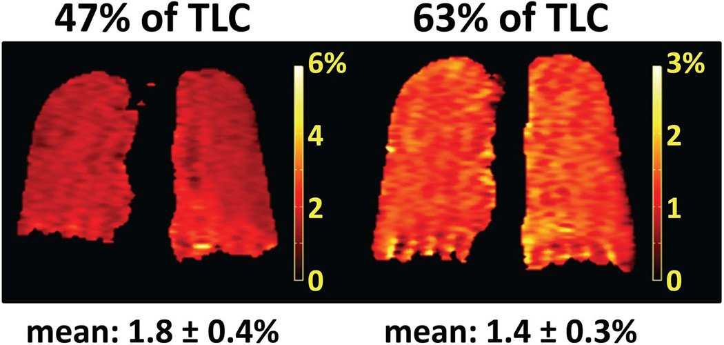
Figure 13.
XTC in a healthy human subject. Maps of fractional gas exchange were calculated from single breath-hold XTC acquisitions at 0.2T (25) performed at 47% (left) and 63% (right) of total lung capacity (TLC) in a healthy volunteer. The mean value of the fractional gas exchange decreased from 1.8% (47% of TLC) to 1.4% (63% of TLC) in response to the increase in lung volume. Although the fractional gas exchange appeared fairly uniform across the lung in both cases, summation of the values along the left-right direction revealed an increase in fractional gas exchange of roughly 0.5% from apex to base for 47% of TLC, whereas the corresponding values for 63% of TLC were uniform from apex to base (25). The time delay between applications of high-flip-angle RF pulses to the dissolved-phase compartments was 62 ms; other acquisition parameters are described in ref. (25). Note that the color scale for the map on the left has a maximum of 6%, while that for the map on the right has a maximum of 3%. (Maps of fractional gas exchange reprinted from European Journal of Radiology, 64(3), Patz S, Hersman FW, Muradian I, et al., Hyperpolarized 129Xe MRI: a viable functional lung imaging modality?, 335–344, Copyright 2007, with permission from Elsevier.)

