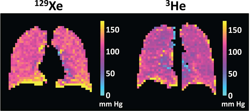
Figure 9.
Comparison of oxygen-concentration imaging using 129Xe and 3He. Coronal maps of the partial pressure of oxygen (pO2) were obtained in healthy subjects using either 129Xe (left) or 3He (right). For both gases, the pO2 values are generally uniform and in a range consistent with normal lung physiology, except for artifactually high values at the base of the lung caused by slight motion of the diaphragm during the acquisitions. Both images were obtained using a gradient-echo-based, cardiac-triggered, short-breath-hold implementation of pO2 mapping (101). (129Xe pO2 map is from healthy subject #1 in ref. (101).)

