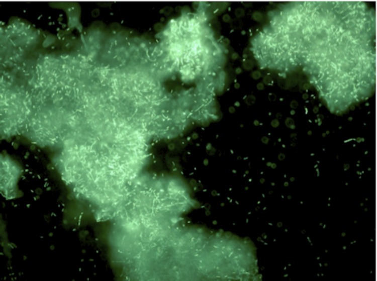
Figure 2C.
Micrograph of Paenibacillus strain Y412MC10 cells showing individual cells and clumps of cells. Cells were grown in YTP-2 media for 18 hours at 37°C and 200 rpm. An aliquot was removed and stained using a 5 μM solution of SYTO® 9 fluorescent stain in sterile water (Molecular Probes). Dark field fluorescence microscopy was performed using a Nikon Eclipse TE2000-S epifluorescence microscope at 200× magnification using a high-pressure Hg light source and 500 nm emission filter.

