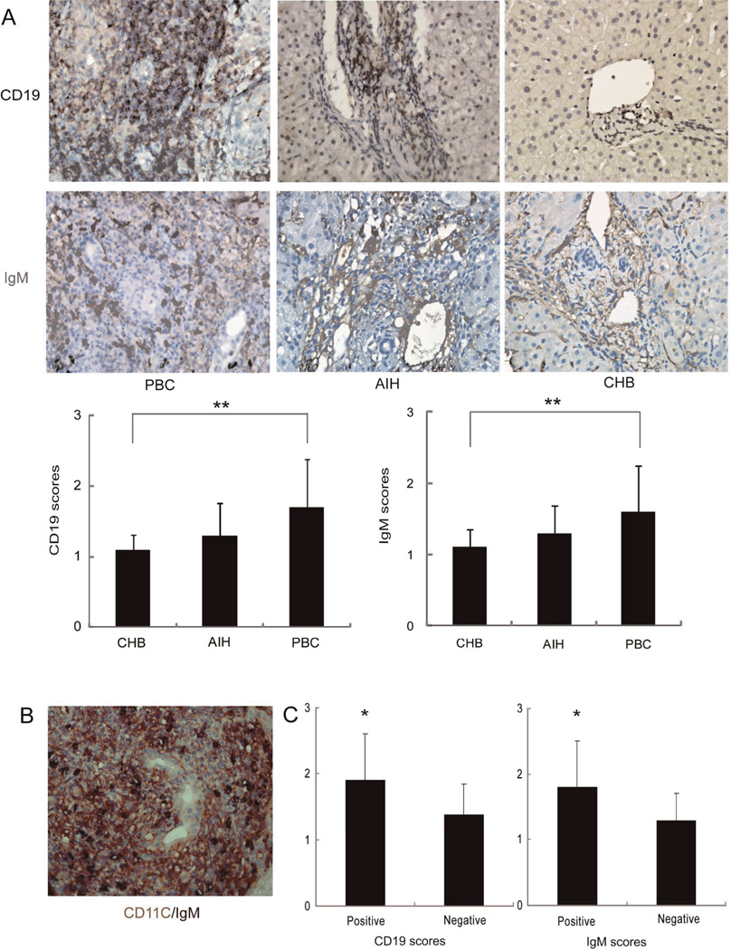
Figure 4.
Presence of B cells and IgM in PBC granulomas. Panel A: Representative immunohistochemical staining for CD19 (magnification 400×) and IgM (magnification 400×) in liver samples from PBC (n=51), AIH (n=37) and CHB (n=30). Five fields were randomly selected for observation in every sample and CD19 and IgM-positive areas scored on a 0-4 point scale for comparison between diseases (** p<0.01, respectively). Panel B: double immunohistochemical staining performed for CD11c (Red) and IgM (Black). Panel C: CD19 and IgM scores in the portal tracts were compared between granuloma positive (n=29) and granuloma negative (n=22) patients with PBC (* p<0.05).

