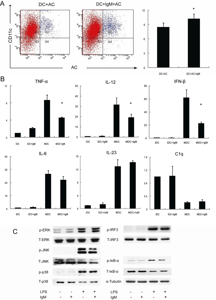
Figure 6.
IgM inhibition of DC phagocytosis. Panel A: myeloid DC were pretreated with IgM (20 µg/ml) then incubated with apoptotic cells (CSFE stained). The percentages of CD11c and CSFE double positive cells were compared between groups treated with or without IgM (* p<0.05). Panel B: DC were pretreated with IgM (20 µg/ml) for 1 hour, then using LPS stimulated for 6 hours. RT-PCR was used to analyze the expression of IL-12p40, IL-6, IL-23p19, TNF-α, IFN-β, C1q, and β-actin (* p<0.05). Panel C: DC were pretreated with IgM (20 µg/ml) for an hour, then total protein was extracted from DCs after stimulated with LPS (1µg/ml) for 30 min. Immunoblots were performed with primary antibodies against phosphorylated or total ERK1/2, JNK p38, IKB-α, IRF3. α-Tubulin expression was determined as the loading control.

