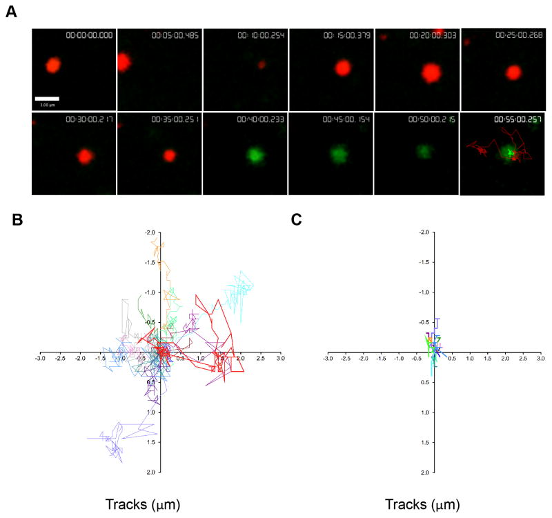
Figure 3. Lytic granules navigate the cell cortex prior to degranulation.
NK92 cells expressing pHlourin-LAMP1 (green) were loaded with LysoTracker Red (red) and activated by immobilized antibody to NKp30 and CD18. Cells were imaged by TIRFm at 6 frames per minute for 60–80 minutes. A. Representative NK92 lytic granule is shown at 5-minute intervals following 10 minutes of cell contact with the activating surface. Granules were tracked using Volocity software prior to and following degranulation as described in Materials and Methods. Pre-degranulation LysoTracker Red (red) and post-degranulation pHluorin-LAMP1 (green) tracks are shown in the final 55-minute image. Scale bar=1 μm. B. Overlay of LysoTracker Red tracks of 14 pre-degranulation events over 4 separate experiments. Lytic granule track from (A) shown in bold (red). C. Overlay of pHluorin-LAMP1 tracks of corresponding degranulation events. Lytic granule track from (A) shown in bold (green).

