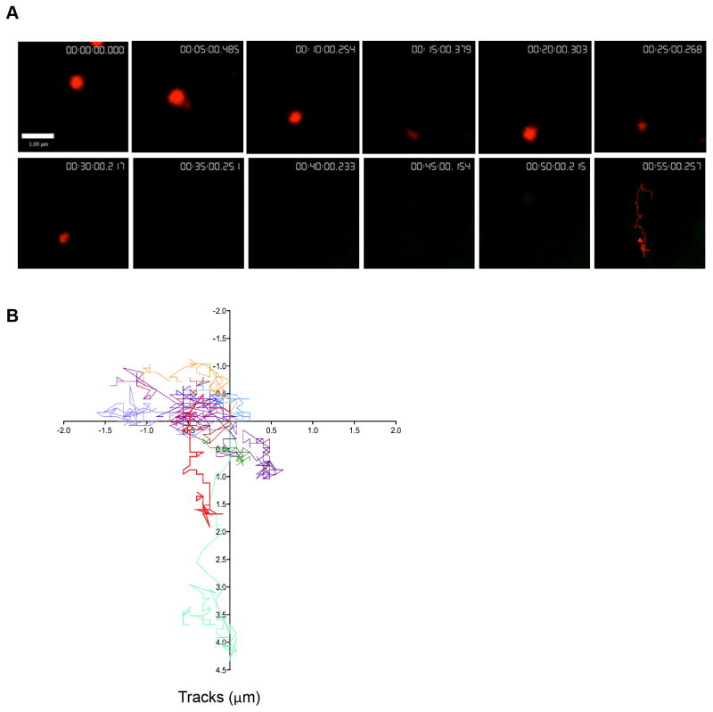
Figure 5. Lytic granules that do not degranulate show normal synaptic motility.
Lytic granules demonstrating no observed degranulation were analyzed. A. Representative NK92 lytic granule cropped from image sequence is shown at 5-minute intervals following 10 minutes of contact. Lysotracker Red (red) track denoting all observed timepoints is shown in final 55-minute image. Scale bar=1 μm. B. Overlay of LysoTracker Red tracks of 10 lytic granules over 4 separate experiments. Representative granule track from (A) is depicted in bold (red).

