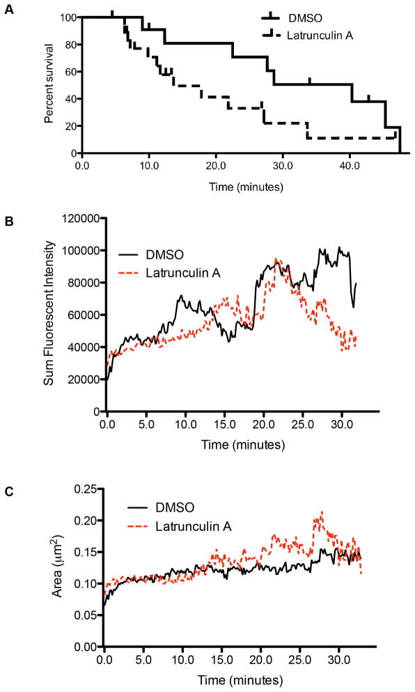
Figure 8. The synaptic actin network is required for the persistence of degranulation.
A. The lifetime of lytic granules post-degranulation in NK cells treated with LatA or DMSO control. Time points reflect amount of time elapsed post-degranulation (marked by the appearance of LAMP1-phluorin) with vertical drops indicating disappearance of the granule from TIRF field. Vertical ticks indicate granules persisting to the end of the imaging sequence. B. Area of the observed lytic granules in cells treated with DMSO (solid black), or LatA (dashed red). C. Sum fluorescent intensity of lytic granules in NK92 cells expressing pHluorin-LAMP1. Cells were treated with DMSO (solid black), or LatA (dashed red) as per Figure 7. Note that sum fluorescent intensity is a function of both the area and mean fluorescent intensity of a lytic granule. Granule boundaries were defined using fluorescent intensity with 3 SD above background as a cutoff. Results shown are from 4 independent experiments.

