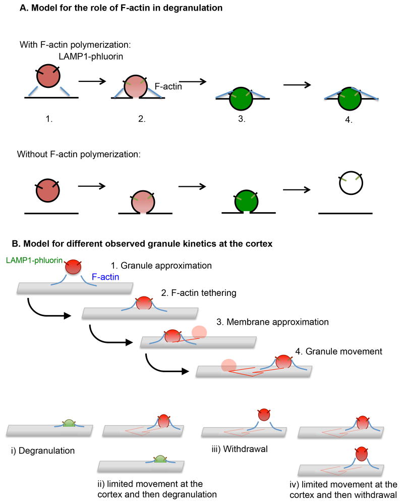
Figure 9. Models for the role of F-actin in granule persistence and the varying behavior of lytic granules at the NK cell cortex.
A) A LysoTracker Red-loaded LAMP1-pHlourin expressing granule is depicted approaching within cell cortex nearing a region within the F-actin network suitable for membrane access (as previously demonstrated (5, 7) (1). As docking and fusion occurs (2), F-actin acts as a tether to help anchor the granule at the membrane, although in both cases fusion results in the activation of LAMP1-phluorin and the subsequent appearance of green fluorescence. In addition, actin reorganization is likely to act in the generation of force to aid in the focused expulsion of granule contents (as supported by greater area*intensity of pHluorin-LAMP1 in control-compared to LatA-treated cells) (3) and the continued persistence of the degranulating granule at the cortex which we would propose is a feature of the interaction of the granule with the local F-actin network (4). B) A LysoTracker Red loaded LAMP1-pHlourin expressing granule approaches the cell membrane and docks with the aid of F-actin tethering (1, 2). This is followed by the approximation of the granule to the cell cortex, movement, then one of the outcomes depicted below. i) immediate exocytosis, ii) limited movement and exocytosis, iii) immediate withdrawal from the cortex or iv) limited movement before withdrawal.

