
Figure 1.
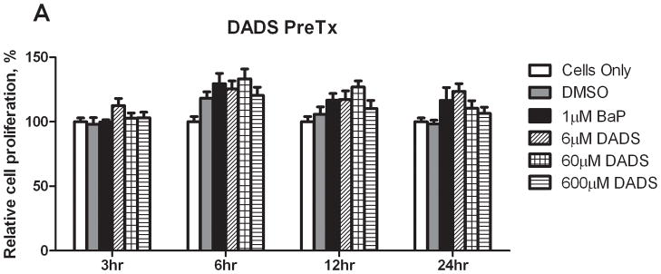
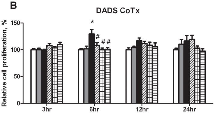
Relative cell proliferation of cells treated with BaP and DADS. MCF-10A cells were pretreated with DADS for four hours followed by the addition of 1 μM BaP (A) or treated concomitantly with 1 μM BaP and DADS (B). To determine cell viability, the MTS solution was applied to the cells for 2–3 hours and analyzed at an absorbance of 490nm. The absorbance of each treatment group was normalized relative to the cells only control for 100% cell viability. The graphs represent the average relative cell proliferation in quadruplicate for N=3, +/− the SEM (* and # indicate a P<0.05 significant difference from the DMSO control and the 1 μM BaP only control, respectively).