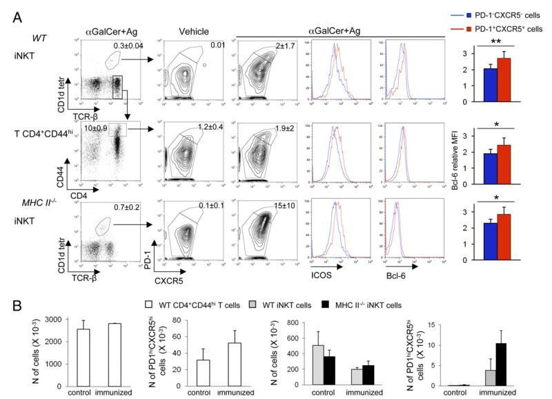
FIGURE 3.
Immunization of MHC II−/− mice with αGalCer+Ag induces iNKTFH cell differentiation. (A) Spleen cells from WT or MHC II−/− mice obtained 7 d after immunization with αGalCer+Ags mix or with the vehicle alone were stained to assess CXCR5, PD-1, ICOS, and Bcl-6 upregulation among CD1d-tetramer+TCRβ+ iNKT cells or TCRβ+CD4+CD44hi Ag-experienced T cells. Numbers in dot plots quadrants indicate the mean percentage ± SD of the cells included in the gates. The histograms on the right show the increase in intracellular Bcl-6 expression between nonfollicular helper (PD-1− CXCR5−) and follicular helper (PD-1+CXCR5+) iNKT or T cells, quantified as the ratio between the mean fluorescence intensity (MFI) of cells stained with anti–Bcl-6 mAb and the MFI of cells stained with isotype controls. (B) Absolute numbers of total T CD4+CD44hi, T CD4+CD44hiPD-1hiCXCR5hi TFH, total iNKT cells, and PD-1hiCXCR5hi iNKTFH in spleens of WT and MHC II−/− mice 7 d after immunization with vehicle or αGalCer+Ags. Numbers were obtained by multiplying the percentage of the relevant target cells by the total number of splenic lymphocytes obtained from the immunized mice. One of three comparable experiments, each performed with three to four mice/group, is shown. *p ≤ 0.05, **p ≤ 0.01.

