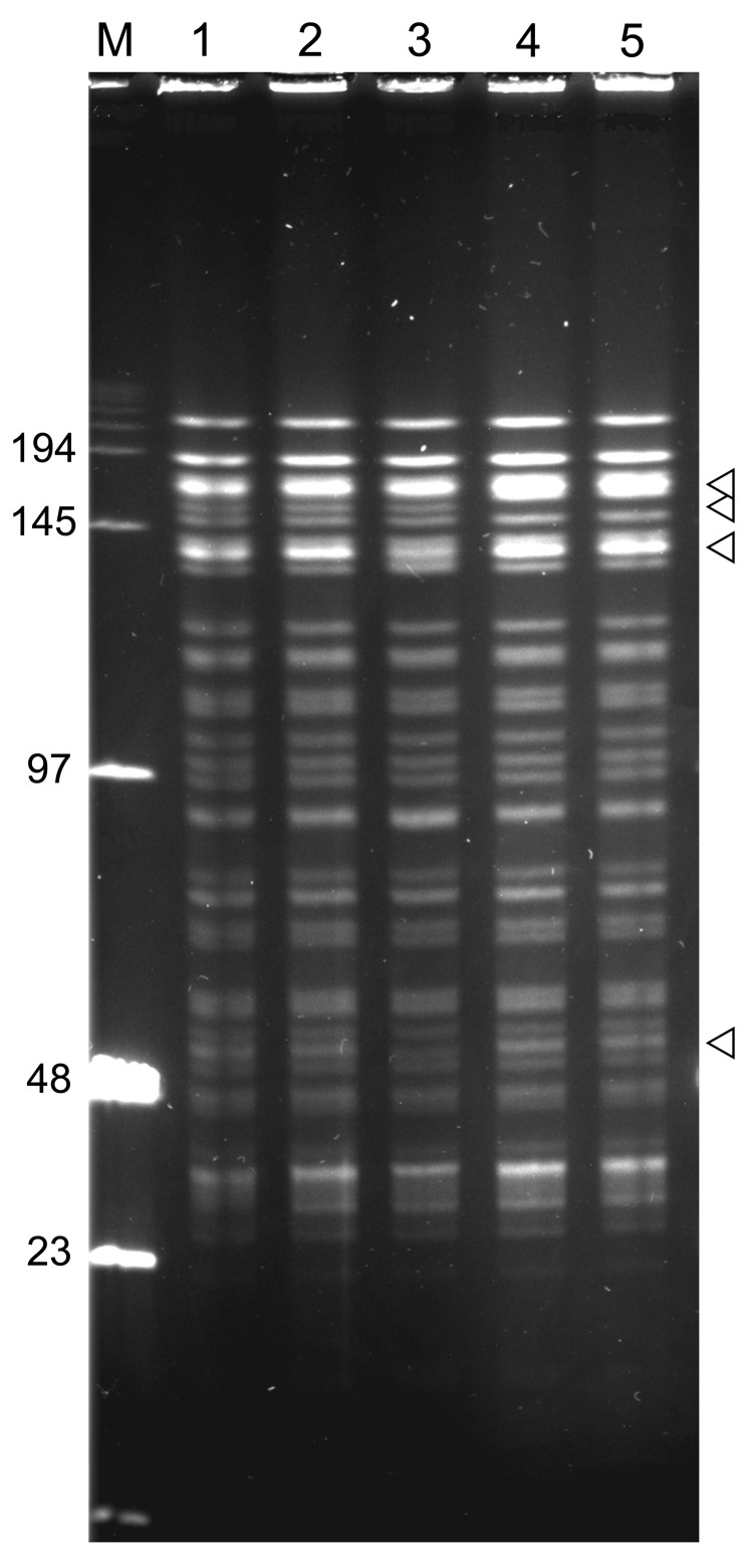
Figure 5.
NotI pulsed-field gel electrophoresis patterns of Yersinia pestis isolates from plague outbreak in Algeria, 2009. Lane M, low-range DNA marker (New England Biolabs, Ipswich, MA, USA); lane 1, IP1860 (Kehailia); lane 2, IP1861 (Hama Ali); lane 3, IP1862 (Hamoul); lane 4, IP1863 (Ain Temouchent); lane 5, IP1864 (Ain Temouchent). Values on the left are in kilobases. Arrowheads indicate positions of variable bands.

