Figure 5. .
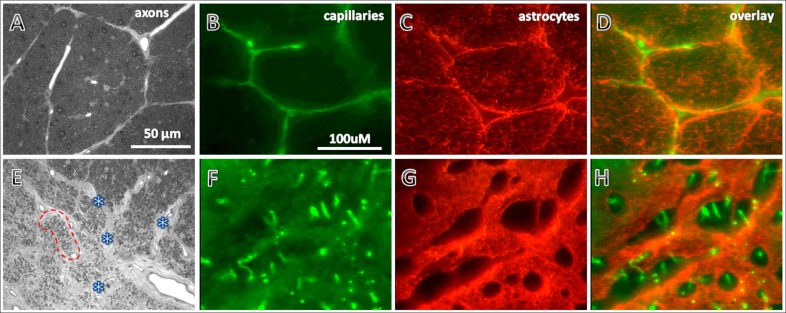
Micrographs show histological observations in one representative BOA eye (bottom panels) compared with a representative control eye (top panels). Axons (A, E) were stained with toluidine blue. Capillaries (B, F) and astrocytes (C, G) were immunohistochemically stained with endothelium markers CD31 and GFAP, respectively, in 50 μm thick sections. Compared with those in the normal eye (A), axons in the damaged area of the BOA eye (E, lower left area) were so scarce that the corresponding fascicles were shrunken and collapsed (red dotted lines encircle one of the collapsed fascicles). The connective tissues in the septa became thickened (marked with asterisks). These changes in the BOA eye led to an appearance of higher capillary density (F) compared with normal eyes (B). In the same area, the GFAP immunoreactivity in the BOA eye was significantly increased (G) compared with the normal eye (C). Overlay of capillaries (D) and astrocytes (H) in the normal and BOA eyes, respectively. Scale bars: (A, E) 50 μm; other photographs 100 μm.

