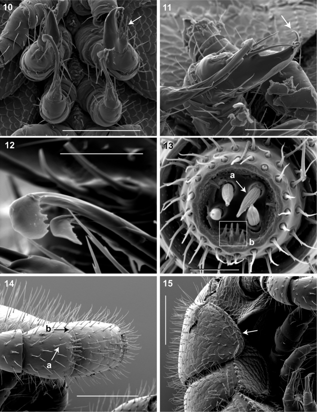
Figures 10–15.
10 Ventral in situ view of gonopods (♂). Arrow, anterior gonopod thick, more robust than posterior gonopod. Scale bar 0.1 mm 11 Medial view of right gonopods (♂). Arrow, Anterior gonopodal apex (podomere 6) shovel-shaped; in repose cupped sheath-like around flagelliform posterior gonopodal apex (podomere 6). Scale bar 0.05 mm. 12 Oblique (right) view of right posterior gonopodal apex (♂). 2 dorsal-most, longest articles laminate distally and recurved laterally, with denticulate posterior margins appearing claw-like. Scale bar 0.02 mm. 13 Antennomere 7 apex (♂). a Four apical cones (AS) oriented in a trapezoidal cluster on 7th antennomere, with longitudinally grooved outer surface and apical circular invagination b Spiniform basiconic sensilla (Bs3) in cluster of 5, oriented apical dorsally on 7th antennomere; tips facing apical cones (on longitudinal axis with Bs2 on antennomeres 5, 6); each sensillum with 2 barbules. Scale bar 0.02 mm.14 Lateral (right) view of right antenna (♂). a Chaetiform sensilla (CS) widely spaced on antennomeres 1-7, each sensillum with 2 or 3 barbules b Trichoid sensilla (TS) oriented apically encircling antennomeres 1–7, lacking barbules. Scale bar 0.1 mm. 15 Lateral (right) view of head, collum and segments 2, 3 (♂).Arrow, collum with carina present on anterolateral margin, appearing scaly. Carina repeated serially on lateral tergal and pleural margins (absent from telson). Scale bar 0.1 mm.

