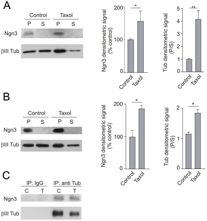
Figure 7. Pharmacological perturbation of the cytoskeleton suggests association of Ngn3 to tubulin and microtubules.
(A) Cultured neurons were untreated or treated with paclitaxel (Taxol; 20 µM) for 40 minutes; then cells were lysated and centrifuged. Proteins present in the precipitate, that includes microtubules and associated proteins (P) and supernatant (S) were analyzed by Western blotting. Ngn3 is enriched in the insoluble fractions and its concentration increases as polymerized tubulin does. (B) Embryonic mouse brains were homogenized and a high-speed precipitate was resuspended and divided in two. Aliquots were left untreated or treated with 20 µM paclitaxel plus 1 mM GTP for 40 minutes at room temperature. Microtubular fraction sedimented by centrifugation (P) and supernatant (S) were analyzed by Western blotting. (C) The supernatants (S) of control (C) and paclitaxel (T) treated aliquots were immunoprecipitated (IP) with anti-βIII-tubulin antibody (or IgG control) to determine the interaction of Ngn3 with soluble tubulin. Precipitates were analyzed by Western blotting with anti-Ngn3 antibody. Graphs show the quantification of densitometry. Error bars show the mean+s.e.m. of three experiments. Significance levels were determined for the data sets connected by horizontal lines using the Student’s t-test. * p<0.05, **p<0.01.

