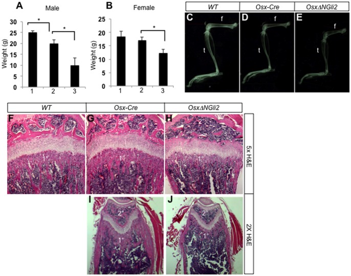
Figure 3. Growth defects and osteopenia in OsxΔNGli2 mice at 6 weeks of age.
(A–B) Body weights of males (A) or females (B) with indicated genotypes. 1: wild type; 2: Osx-Cre; 3: OsxΔNGli2. n = 3; *: p<0.05; error bar: STDEV. (C–E) X-ray radiography for the hindlimbs of wild type (C), Osx-Cre (D) and OsxΔNGli2 (E) male mice at 6 weeks of age. f: femur; t: tibia. (F–H) H&E staining of sections from the proximal ends of tibias in 6-week-old male mice. (I, J) H&E staining of sections from the distal ends of femurs in 6-week-old male mice. 1°: primary ossification center; 2°: secondary ossification center. Note decrease in bone in both centers of OsxΔNGli2 mice.

