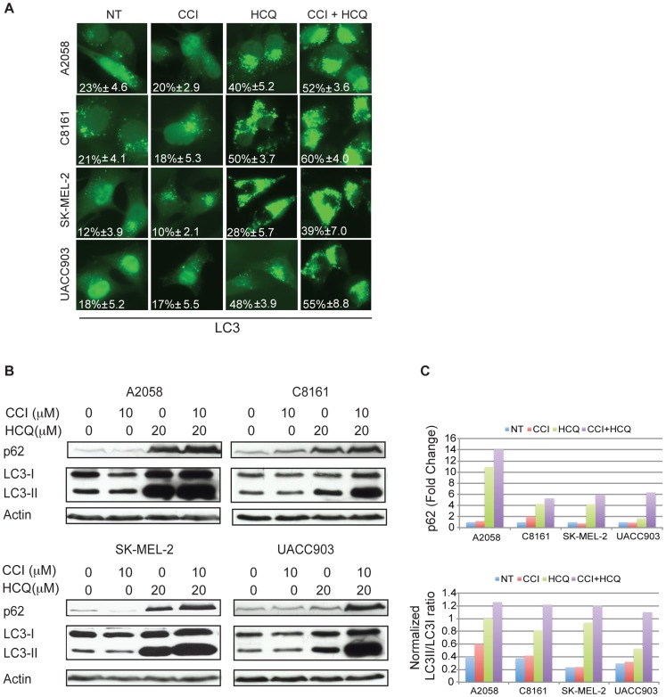
Figure 2. Autophagic flux is induced by CCI-779 and blocked by HCQ.
(A) Melanoma cell lines transiently transfected with an EGFP-LC3-expressing plasmid were seeded on coverslips and treated with 10 µM of CCI-779 and 20 µM of HCQ, alone and in combination for 24 hours. Autophagosomes were visualized by the presence of LC3 puncta. The drug combination treatment showed more autophagosome accumulation than either single agent alone. The percentages of cells showing LC3 puncta (mean ± SD) are indicated. (B) Western blot showed that the combination treatment resulted in considerable increases in LC3-II and increases in p62 protein levels compared to treatment with single agent HCQ alone, indicating the blockade of autophagic flux. (C) Quantitation of actin-normalized changes in p62 (upper panel) and the ratio of LC3-II/LC3-I (lower panel) after single and combination treatment in comparison to the untreated control.

