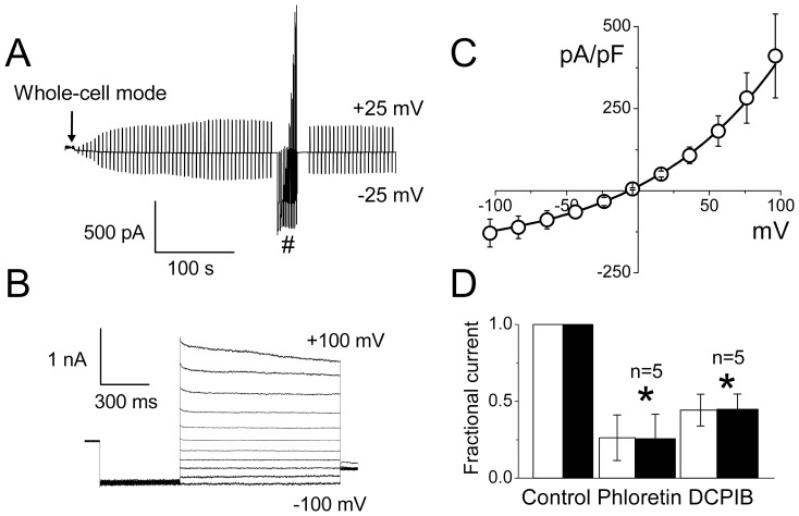
Figure 5. Whole-cell macroscopic currents activated by cell swelling in rat thymocytes.
(A) Time course of whole-cell current activation in response to cell swelling. Currents were elicited by application of alternating pulses from 0 to ±25 mV (every 5 s). (B) Representative traces of current responses recorded at the steady-state in (A) marked with # symbol. The holding potential was 0 mV; after a pre-pulse to −100 mV (500 ms), currents were elicited by application of step pulses (1000 ms) from −100 to +100 mV in 20-mV increments. (C) Instantaneous I-V relationship measured at the beginning of test pulses from recordings similar to those shown in (B). (D) Effects of VSOR channel blockers, phloretin (200 µM) and DCPIB (10 µM), on the whole-cell macroscopic currents activated by cell swelling in rat thymocytes. Open and filled bars represent the fractional currents measured at +25 mV and −25 mV, respectively. *Significantly different from the control values at P<0.05.

