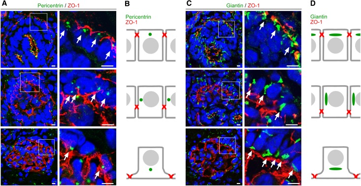
Figure 7.
Translocation of the centrosome and Golgi apparatus during podocyte differentiation. (A) Frozen kidney sections of newborn wild-type rat are stained using antibodies against the centrosome marker protein pericentrin (arrows) and the junction marker ZO-1 and are subjected to confocal laser microscopy (top, comma-shaped body stage; middle, late s-shaped body stage; bottom, immature glomerulus). (B) Schematic illustration of the pericentrin signal during podocyte differentiation. (C) Frozen kidney sections of newborn wild-type rat are stained using antibodies against the Golgi apparatus marker giantin (arrows) and ZO-1. Arrows mark translocation of the Golgi apparatus from apical to basal during podocyte differentiation (top, comma-shaped body stage; middle, late s-shaped body stage/early capillary loop stage; bottom, immature glomerulus). (D) Schematic illustration of the giantin signal during podocyte differentiation. Scale bars, 5 µm.

