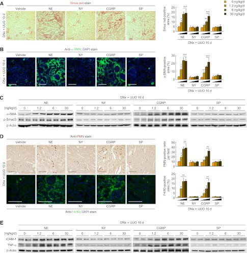
Figure 2.
Norepinephrine and CGRP contribute to kidney fibrosis and inflammation during UUO. Renal denervation (DNx) in left kidneys of male 129S1/SvImJ mice aged 8–10 weeks was carried out; 2 days after the onset, norepinephrine (NE), NY, CGRP, or substance P (SP) (0, 1.2, 6, or 30 ng/kg per day) was continuously infused into kidneys via a mini-osmotic pump, and the left ureters were obstructed for 10 days. (A) Norepinephrine- and CGRP-induced collagen deposition using Sirius red staining in denervated UUO kidneys. The Sirius red–positive area was measured in five randomly chosen high-power (×200) fields per kidney using NIH ImageJ software. (B) Immunohistochemistry of α-SMA in denervated UUO kidneys after treatment. The visible blue color indicates nuclei stained by DAPI. The α-SMA–positive area was measured in five randomly chosen high-power (×200) fields per kidney using NIH ImageJ software. (C) α-SMA and p-Smad3 expression in norepinephrine- or CGRP-treated UUO kidneys using Western blot analysis. Anti–β-actin antibody served as a loading control. (D) Immunohistochemistry of PMN and F4/80 in norepinephrine- or CGRP-treated UUO kidneys. The number of PMN-positive neutrophils and the area of F4/80-positive macrophages were evaluated in five randomly chosen high-power (×200) fields per kidney. (E) ICAM-1 and TNF-α expression using Western blot analysis. Anti–β-actin antibody served as a loading control. n=6 in each group. Scale bar, 50 μm. *P<0.05, **P<0.01, ***P<0.001 versus 0 ng/kg per day. Error bars represent SDs.

