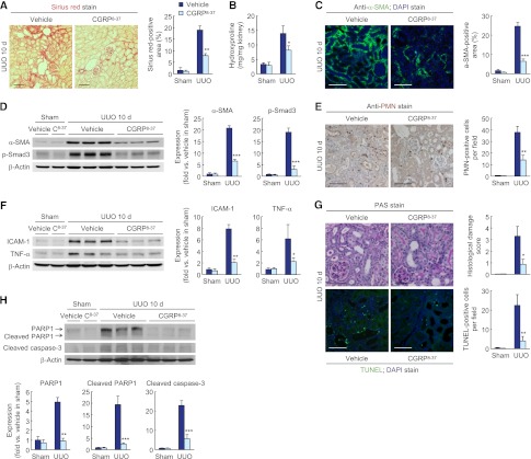
Figure 7.
CGRP inhibition reduces tubulointerstitial fibrosis, inflammation, and tubular cell death during UUO. Male 129S1/SvImJ mice aged 8–10 weeks were given a CGRP receptor antagonist (CGRP8–37 or C8–37; 120 μg/kg per day) or 0.9% saline (vehicle) via an intraperitoneal implantation of the mini-osmotic pump from 24 hours before UUO or sham operation (n=5). (A and B) Collagen deposition was evaluated by Sirius red staining (A) and hydroxyproline measurement (B) in UUO kidneys treated with CGRP receptor antagonist or vehicle. (C) α-SMA expression in UUO kidneys treated with CGRP receptor antagonist or vehicle using immunohistochemistry. The visible blue color indicates nuclei stained by DAPI. (D) α-SMA and p-Smad3 expression using Western blot analysis. Anti–β-actin antibody served as a loading control. (E) Immunohistochemistry of PMN in UUO kidneys treated with CGRP receptor antagonist or vehicle. (F) ICAM-1 and TNF-α expression in UUO kidneys treated with CGRP receptor antagonist or vehicle using Western blot analysis. Anti–β-actin antibody served as a loading control. (G) Histologic damage in UUO kidneys treated with CGRP receptor antagonist or vehicle indicated by PAS staining. TUNEL assay in UUO kidneys treated with CGRP receptor antagonist or vehicle using In Situ Cell Death Detection kit. The visible blue color indicates nuclei stained by DAPI. (H) PARP1, cleaved PARP1, and cleaved caspase-3 expression in UUO kidneys treated with CGRP receptor antagonist or vehicle using Western blot analysis. Anti–β-actin antibody served as a loading control. Scale bar, 50 μm. *P<0.05, **P<0.01, ***P<0.001 versus vehicle. Error bars represent SDs.

