Figure 1.
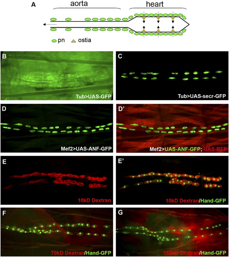
Drosophila pericardial nephrocytes uptake secreted proteins. (A) Schematic drawing of Drosophila heart. pn, pericardial nephrocyte. (B) Larva with Tub-Gal4 and UAS-GFP showed ubiquitous expression of GFP in all tissues. (C) Larvae with Tub-Gal4 and UAS-secr-GFP showed GFP accumulation specifically in nephrocytes. (D and D′) Embryos with Mef2-Gal4 and UAS-ANF-GFP together with UAS-RFP showed specific accumulation of ANF-GFP in pericardial nephrocytes (green) and RFP in muscles (red). (E–G) Larva with Hand-GFP transgene (green) injected with (E and E′) 10-, (F) 70-, or (G) 155-kD fluorescence-labeled Dextran (red) showed size-dependent Dextran uptake, with (E and E′) high-efficient uptake of 10-kD Dextran, (F) less-efficient uptake of 70-kD Dextran, and (G) no uptake of 155-kD Dextran.

