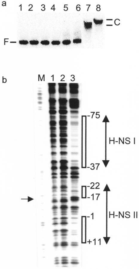
FIG. 4.
Location of H-NS binding sites in PhlyE. (a) H-NS binds at PhlyE. Lane 1, no protein; lanes 2 to 8, H-NS (0.04, 0.08, 0.12, 0.25, 0.5, 1.0, and 2.0 μM, respectively). The positions of the retarded complexes (C) and free PhlyE DNA (F) are indicated. (b) DNase I footprints of H-NS at PhlyE. Lane M, Maxam and Gilbert G track; lanes 1 and 2, no protein; lane 3, H-NS (1 μM). The regions of H-NS protection (open boxes) and the hypersensitive base (arrow at the left) are indicated. All numbering is relative to the SlyA-associated transcription start site (36).

