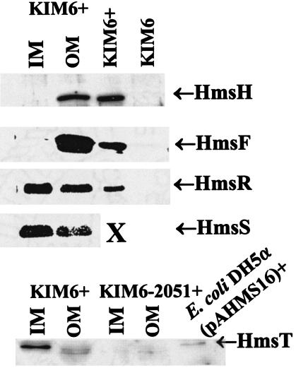
FIG. 2.
Cellular location of Hms proteins. All Hms proteins localized to membrane fractions; consequently, cytoplasmic and periplasmic fractions are not shown. KIM6+, E. coli DH5α(pAHMS16), and KIM6 lanes contain whole-cell extracts used as positive and negative controls. X indicates that unfractionated KIM6+ cell extracts were not tested with antibody against HmsS.

