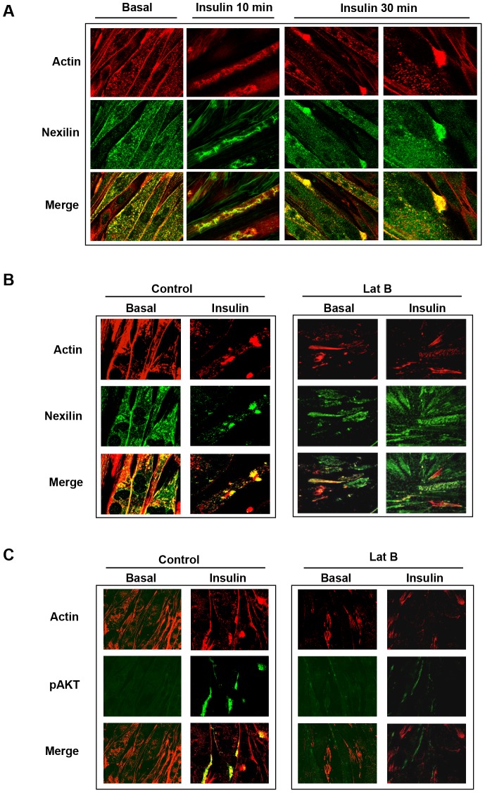
Figure 2. Spatial distribution of nexilin in L6 skeletal muscle cells.
A) L6 myotubes were serum starved (basal) or stimulated with 100 nM insulin as indicated and then fixed, permeabilized and incubated with anti-nexilin abs, Cy5-conjugated secondary antibodies (green) and rhodamine-phalloidin (red). Images were obtained on a Zeiss LSM510 laser scanning confocal microscope; B) Serum depleted L6 myotubes were pre-incubated with or without Latrunculin B (LatB) and subsequently stimulated with 100 nM insulin for 30 minutes. Cells were stained as in A); C) L6 myotubes were treated as in B) and processed for visualization using phospho-Ser473 Akt abs (green) and rhodamine-phalloidin (red).

