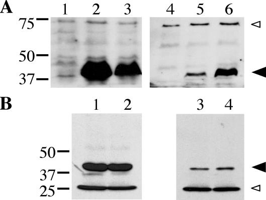
FIG. 7.
Subcellular localization of DotB and DotB K162Q. (A) Total protein (lanes 1 to 3) and total membrane protein (lanes 4 to 6) fractions were taken from three L. pneumophila strains and were subjected to DotB Western blotting. Strains included the ΔdotB strain JV918 with empty vector pJB908 (lanes 1 and 4), JV918 with the DotB complementing clone pJB1153 (lanes 2 and 5), and JV918 with the DotB K162Q clone pJB1568 (lanes 3 and 6). The molecular masses of relevant markers (in kilodaltons) are on the left. (B) Total protein (lanes 1 and 2) and total membrane protein (lanes 3 and 4) fractions were taken from two E. coli strains and were used in DotB Western blotting. Strains included XL1-Blue with the DotB complementing clone pJB1153 (lanes 1 and 3) and XL1-Blue with the DotB K162Q clone pJB1565 (lanes 2 and 4). (A and B) Large arrowhead, DotB; small arrowhead, cross-reacting band that confirms overall sample equivalence. The molecular masses of relevant markers (in kilodaltons) are on the left. Results are representative of several experiments.

