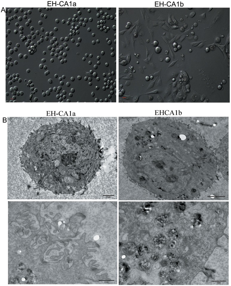
Figure 1. Morphology of EH-CA1a cells and EH-CA1b cells.
(A). EH-CA1a cells are small, round, and grew in suspension, whereas EH-CA1b cells are relatively large, spindle-shaped, and grew as a monolayer (B) Transmission electron microscopy of EH-CA1a and EH-CA1b cell lines (TEM ×7500). Compared with EH-CA1b cells, EH-CA1a cells EH-CA1a cells have more microvillus on the cell surface.

