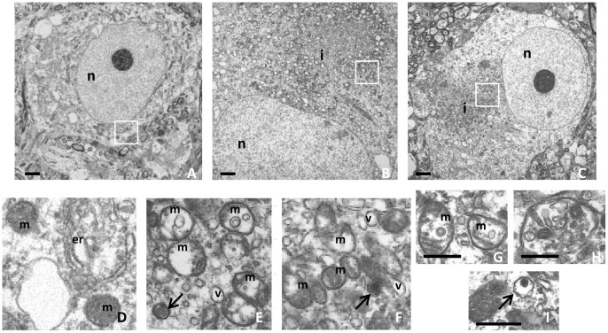
Figure 3. The absence of α-synuclein does not modify the ultrastructure of PB-like inclusions in mouse SNpc neurones.
Representative electron micrographs of control (A; Psmc1 fl/fl;TH Wt) and 26S proteasome-depleted SNpc neurones with (B; Psmc1 fl/fl;TH Cre;Snca +/+) and without (C; Psmc1 fl/fl;TH Cre;Snca −/−) α-synuclein. Enlarged views of the boxed areas are shown in D-F respectively. The inclusions contain mainly abnormal mitochondria (E-G; m) interspersed with numerous small vesicles (E and F; v). Autophagosome-like structures containing electron-dense material (E, F and I; arrows) as well as recognizable cytosolic elements including mitochondria (H) are present. n, nucleus; i, PB-like inclusion; m, mitochondria; v, vesicle and er, endoplasmic reticulum. Scale bar, 500 nm. Psmc1 fl/fl;TH Cre;Snca +/+ (G and H); Psmc1 fl/fl;TH Cre;Snca −/− (I).

