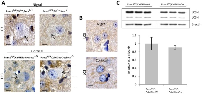
Figure 4. Autophagy is not activated in 26S proteasome-depleted mouse neurones.
(A) Absence of LC3 immunoreactivity in representative neurones of the SNpc (Psmc1 fl/fl;TH Cre, nigral) and cortex (Psmc1 fl/fl;CaMKIIα-Cre, cortical) in the presence (Snca +/+) and absence (Snca −/−) of α-synuclein. The arrows indicate PB-like inclusions. Scale bar, 10 µm. (B) The normal pattern of LC3 in nigral and cortical neurones shows a fine punctate cytoplasmic staining. Neurones from Psmc1 fl/fl;TH Wt;Snca +/+ (nigral) and Psmc1 fl/fl;CaMKIIα-Wt;Snca +/+ (cortical) mice are shown, but the pattern of LC3 immunostaining was similar in the absence of α-synuclein. Scale bar, 10 µm. (C) Representative Western blot of LC3-I and LC3-II in total cortical homogenates from control (Psmc1 fl/fl;CaMKIIα-Wt) and 26S proteasome-depleted (Psmc1 fl/fl;CaMKIIα-Cre) mice. Graph depicts LC3-II levels normalized to β-actin. n = 4, no significant difference. Error bars indicate SEM.

