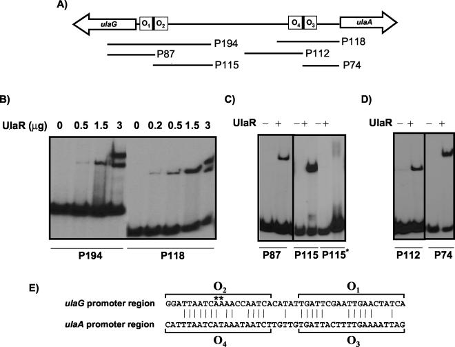
FIG. 4.
Binding of UlaR to different fragments of the ulaG-ulaA intergenic region. (A) Diagram of ulaG-ulaA intergenic region; the fragments used as probes after 32P labeling are indicated by lines. (B) Binding of UlaR to probes P194 and P118. (C) Binding of UlaR added (+) at 1.5 μg to probes P87, P115, and the mutated P115 (P115*). (D) Binding of UlaR added at 1.5 μg to probes P112 and P74. (E) Alignment of the nucleotide sequences (written in the direction of transcription of the ulaG and ulaA genes) of the UlaR binding sites present in fragments P194 and P118. The vertical lines indicate the conserved residues. The nucleotides forming the proposed operator sites are indicated by brackets. Nucleotide changes of the mutation present in P115* abolishing UlaR binding are shown by asterisks.

