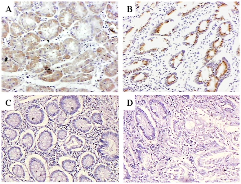
Figure 5. Representative immunohistochemical staining in serial sections of poorly differentiated adenocarcinoma of the stomach from patients positive for AFP (magnification ×100).
(A) Immunostaining for AFP. (B) Strong STAT3 immunostaining with brown granular deposits in the cytoplasm and nuclei. (C) Negative control immunohistochemical staining for AFP. (D) Negative control immunohistochemical staining for STAT3.

