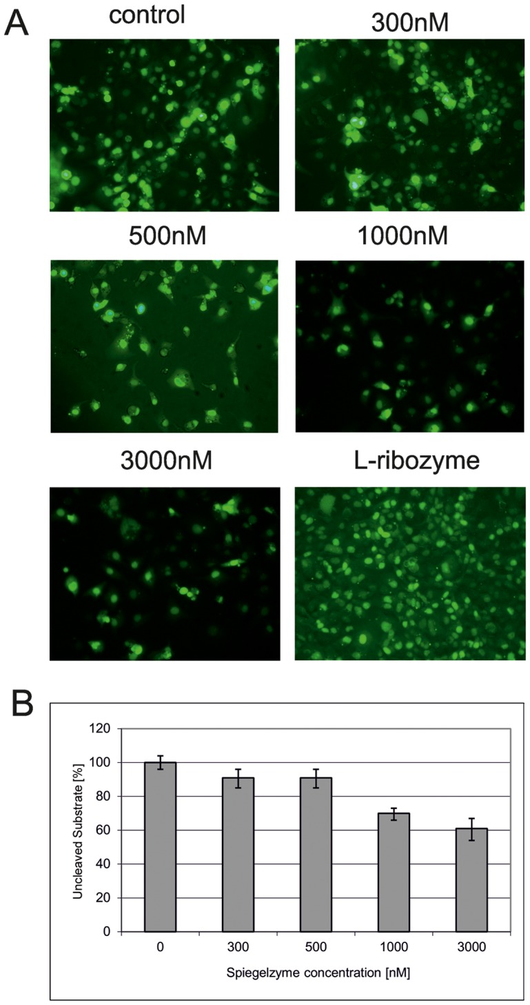
Figure 5. Hammerhead Spiegelzyme Activities in COS-7 cells.

(A) Cells were transfected with 5′-fluorescein labeled L-RNA-1 substrates. Prior to the application of the HH Spiegelzyme, the cells were washed to remove the untransfected substrate from the medium. Then the cells were transfected with 300, 500, 1000 and 3000 nM of the Spiegelzyme. The control transfection was done only with the substrate. To check for the ability of the Spiegelzyme to cross the membrane, the cells were transfected with the fluorescein labeled Spiegelzyme (bottom row, right panel). The microscopic images were taken prior to RNA isolation after washing with PBS buffer. (B) The percentage of uncleaved substrate in the L-RNA-1 isolated from cells transfected with the Spiegelzyme. After incubation for 24 hours, L-RNA was isolated from harvested cells using TRIZOL (Ambion) according to the manufacturer’s protocol. L-RNA-1 was separated by 20% PAGE with 8 M urea and the amount of substrate was determined by the fluorescence intensity using Fuji Film FLA 5100 phosphoimager.
