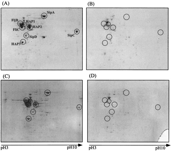
FIG. 6.
Two-dimensional gel electrophoresis patterns of proteins secreted into the medium by S. enterica serovar Typhimurium strains χ3306 (wild type) grown at 30°C (A) or 37°C (C), CS2021 (dnaK::Cm) grown at 30°C (B), and CS2069 (dnaK::Cm, Ts+ suppressor) grown at 37°C (D). Protein spots enclosed in circles were analyzed by mass spectrometry as described in Materials and Methods and are interpreted in the text.

