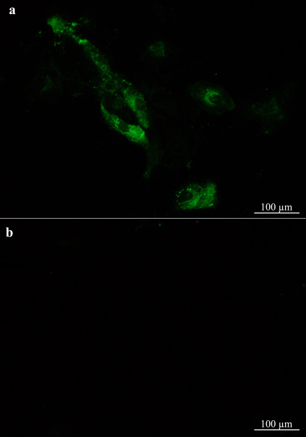
Figure 4.
Localisation of ISAV in Atlantic salmon primary gill epithelial cells. Infected cells (a) fixed at 4 day p.i. and subjected to immunofluorescence (IFAT) staining (green fluorescent staining) with mouse monoclonal antibody directed against the ISAV nucleoprotein. No viral antigen was detected in mock-infected cells (b).

