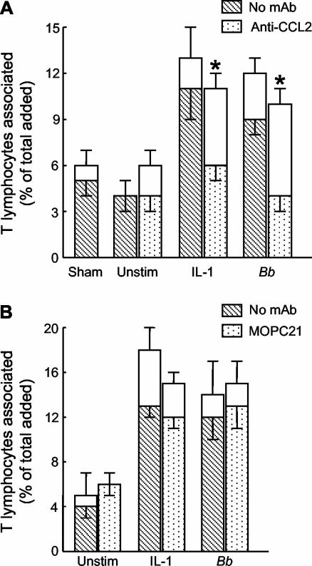
FIG. 1.
Enhanced migration of T lymphocytes across stimulated endothelium is dependent on CCL2 (MCP-1). HUVEC were left unstimulated (Unstim), treated with a sham preparation of bacteria, or stimulated with 1 U of IL-1β per ml or B. burgdorferi (Bb) at 10 spirochetes per endothelial cell for 24 h. During the last 4 h of stimulation, 25 μg of MAb to CCL2 per ml (A), 30 μg of the isotype-matched control MAb MOPC21 per ml (B), or an equal volume of phosphate-buffered saline (No MAb) was added. T lymphocytes (5 × 10⁵ cells/cm2) were then added to the endothelium for 4 h in the presence of additional MAb. The total height of each bar represents the percentage of added T cells that became associated with the cultures. The lower (patterned) portion of each bar represents the percentage of cells that migrated beneath the endothelium; the upper (unfilled) portion illustrates the percentage that was adherent to the apical surface of the HUVEC. Bars represent the means ± standard deviations of three to four replicate samples. For A, data shown are representative of two separate experiments. Asterisks denote significant reductions in migration compared to control assays performed in the absence of MAb (P < 0.001).

