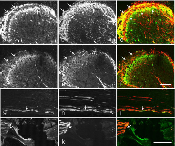
Figure 6.
Scgn-LI in mouse spinal cord and sciatic nerve after rhizotomy or nerve ligation. (a-f) Immunofluorescence micrographs of sections incubated with antiserum against Scgn- (a,c,d,f) and CGRP- (b,c,e,f) LIs in lumbar spinal cord 10 days after unilateral, dorsal rhizotomy (d-f). (a-f) On the contralateral side both Scgn- and CGRP-LIs are strong in the superficial dorsal horn, the former displaying two bands (arrows in a, c), the latter filling out layers I and outer II (arrows in b,c). The merged micrograph (c) shows that the deeper band of Scgn-LI, consisting mainly of cell bodies, essentially remains in inner lamina II, without much overlap with the CGRP-LI (d-f). There is a strong ipsilateral decrease of both Scgn-LI (cf. d, f with a, c; arrows) and CGRP-LI (cf. e, f with b, c), compared to a contralateral side (a-c). (g-i) Scgn-LI is weekly (arrows in g, i), and CGRP more extensively (arrow in h, i) expressed in the control sciatic nerve, and both accumulate, on the proximal side of a-10-hour ligation (arrows in j, k). Double-staining shown in merged color micrographs indicates possible coexistence (arrows in i, l). Scale bars indicate 100 μm (a-f; g-l).

