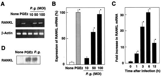
FIG. 2.
Expression of RANKL mRNA by osteoblasts infected with P. gingivalis ATCC 33277. (A) RT-PCR analysis of RANKL. Mouse osteoblasts were cultured in the absence of the stimulant (None) or in the presence of PGE2 (2 μM) for 6 h at 37°C. Other osteoblast cultures were infected with P. gingivalis (P.g.) at MOIs of 10, 50, and 100 for 6 h at 37°C. Total RNA was extracted, reverse transcribed, and subjected to RT-PCR analysis for RANKL and β-actin. (B) Quantification of RANKL by real-time PCR. Cells were cultured in the absence of the stimulant (None) or in the presence of PGE2 for 6 h at 37°C. Other osteoblast cultures were infected with P. gingivalis (P.g.) at MOIs of 10, 50, or 100 for 6 h. RNA was extracted, reverse transcribed, and subjected to real-time PCR. Relative mRNA levels are expressed as percentages of the mRNA level in osteoblasts stimulated with PGE2. (C) Time course of RANKL expression. Osteoblasts were infected with P. gingivalis (MOI = 100) for 1, 3, 6, or 12 h at 37°C. Relative mRNA levels are expressed as the fold increases compared to the RANKL mRNA level in unstimulated cells. The values shown are means ± standard deviations for triplicate assays. An asterisk indicates that the P value was <0.05 in comparison with the RANKL mRNA level in cells cultured in the absence of the stimulant (None). (D) Ligand precipitation of RANKL expressed on the infected osteoblasts. Mouse osteoblasts were cultured in the absence of stimulant (None) or in the presence of PGE2 for 48 h in αMEM containing 10% fetal calf serum. Other cultures of osteoblasts were infected with P. gingivalis (P.g.; MOI = 100) and cultured for 48 h. Cell-associated RANKL was precipitated with RANK-immobilized beads and subjected to SDS-PAGE and Western blotting with anti-RANKL antibodies.

