
Figure 2. Characterization of Homo- and Heterodimeric Structure by Gel Electrophoresis.
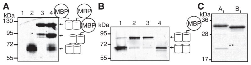
The contrast of each whole image has been adjusted to emphasize weak bands.
(A) Validation of the SDS-PAGE electrophoretic mobility shift screen for pairs of protein variants that exhibit heterodimeric character. Proteins were loaded without boiling and were detected by imaging the red fluorescence. Lane 1, uninduced culture; lane 2, dTomato; lane 3, MBP-dTomato; lane 4, coexpressed dTomato and MBP-dTomato. Asterisk (*) indicates a species resulting from proteolysis of one dTomato-MBP linker.
(B) Red fluorescence image of a gel used for PAGE analysis of four representative variants analyzed during screening (numbered 1–4).
(C) Coomassie-stained gel of purified A1 and B1 variants. Samples were boiled in sample buffer prior to electrophoresis. The double asterisk (**) indicates the 19 kDa product of chromophore hydrolysis routinely observed for RFPs (Gross et al., 2000). The 11 kDa fragment was not observed.