
Figure 6. Ganglion cell axonal ribbon targets.
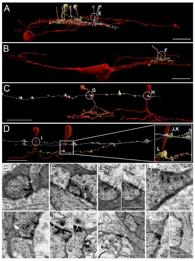
A-D. Renderings of five CBb classes forming axonal ribbons onto multiple ganglion cell classes, vertical orientation. Circles indicate location of synapses shown in E-L. Scale bars (A-B), 25 μm; scale bars (C-D), 20 μm. E-L. TEM of synapses indicated in A-D. White arrows indicate synapse directionality. GC, ganglion cell; WF BC, wide field bipolar cell; AC, amacrine cell; r, ribbons; c, cistern; pcd, post-citernal density; scale bars, 0.5 μm;. A,E. CBb4 3116 (left cell of the silver pair that intersect ganglion cell 15796 (red)) forms an axonal single-ribbon dyad with bsdGC 15796 and an unknown cell. CBb4 3116 participates in a chain of seven coupled CBb3s (tan) and CBb4s (silver). The bsdGC 15796 dendritic target of the axonal ribbon abruptly ascends to the OFF IPL where it receives the input before returning to the ON IPL distally (far right of panel A). B,F. CBb5 400 (mustard) forms an axonal multi-ribbon dyad with ON-OFF ganglion cell 5118 (red) and an unknown cell. C,G,H. CBb5w 6156 (copper) and wide-field cone bipolar cell 5283 (red) converge an axonal single-ribbon monad and axonal multi-ribbon monad, respectively, onto ipRGC 12208 (off white). Note the omega figure in the right subpanel of panel G. Wide-field cone bipolar cell 5283 forms an axonal cistern onto γAC 20537 (not shown in C, see Fig. 8, B-C) in the same plane of section as the four-ribbon axonal monad onto ipRGC 12208. D,I,J,K,L. CBb6 353 (red, left cell) and CBb6 447 (red, right cell) both form multi-ribbon axonal dyads (I,J) onto OFF-layer monostratified ganglion cell 21779 (silver) and another amacrine cell, amacrine cell 22210 (not shown inD for clarity,I) and amacrine cell 32273 (upper bright green cell in D, D inset,J), respectively. Amacrine cell 32273 creates both feedback (J, right subpanel) and feedforward (K) inhibition motifs via conventional synapses onto CBb6 447 and ganglion cell 21779, respectively. CBb6 447 also forms a single-ribbon axonal dyad in the ON IPL onto multistratified ganglion cell process 34336 (beige in D, D inset,L left subpanel) and amacrine cell 34337 (lower bright green cell in D, D inset,L left subpanel). Amacrine cell 34337 forms a conventional synapse onto ganglion cell 34336 (L right subpanel), thus completing a feedforward inhibition motif.