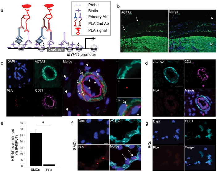
Figure 1. ISH-PLA: a new method of detection of histone modificationsat a single genomic locus in tissuesections.
(a) Schematic of the combined in situ hybridization (ISH) and Proximity Ligation Assay (PLA) method for detecting H3K4dime of the MYH11 promoter. (b) Workflow of the ISH-PLA procedures. (c) Immunostaining of 5 μm-thick sections of human carotid artery for ACTA2 and DAPI (diamidinophenyindole). Two distinct layers are identified: the media (M) and the adventitia (A). Small vessels consisting of ACTA2+ SMCs are visualized within the adventitia (arrows). Scale bar = 100 μm. (d) ISH-PLA in adventitial small arteries (n = 5). PLA amplification (PLA+) is visualized as a red spot localized within nuclei. MYH11 H3K4dime PLA+ signal is restricted to ACTA2+ medial SMCs (arrows) and is absent from CD31+ ECs (stars) as well as adventitial fibroblasts (arrow heads). (i–iii): High magnification with i) ACTA2, ii) PLA and iii) merge.Scale bar = 50 μm. (e)ISH-PLA negative control using an empty vector probe in adventitial vessel of human carotid artery sections. A total absence of PLA amplification demonstrates that hybridization of the biotin-labeled MYH11 probe is required for ISH-PLA amplification. Scale bar = 50 μm (f) Conventional ChIP assays showing enrichment of H3K4dime of MYH11 in SMCs but not in ECs. Mean ± s.d.; n = 3; *P < 0.05. (g) ISH-PLA in SMCs (n = 3) and ECs (n = 3) in vitro. MYH11 H3K4dime PLA amplification is restricted to SMCs.Scale bars = 10 μm.

