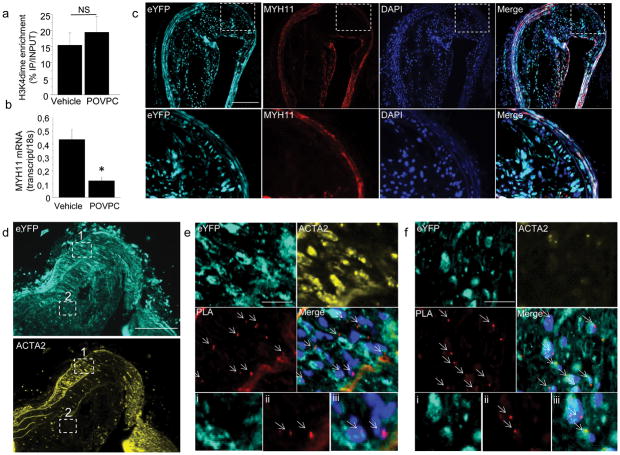
Figure 4. H3K4dime on the MYH11 promoter persists during phenotypic switching in vivo in SMC lineage tracing mice developing atherosclerosis.
(a) ChIP assay performed in cultured SMCs treated with vehicle (DMSO) or POVPC (10 μg/mL, 24h) showing similar H3K4dime enrichment on the MYH11. Mean s.d.; n = 3. (b) mRNA quantification of MYH11 showing POVPC-induced decrease in MYH11 mRNA level compared with vehicle. Mean ± s.d; n = 3; *P <0.01. (c)Identification of modulated SMCs in SMC-eYFP+/+ ApoE−/− mice fed with Western diet for 18 weeks. Brachiocephalic arteries (BCA) sections of these mice were stained with eYFP (cyan) and MYH11 (red). eYFP+ SMCs were identified within the media where they co-express MYH11 and within the atherosclerotic lesion where they lose expression of MYH11. Scale bar = 100 μm. (d) BCA sections of SMC-eYFP+/+ ApoE−/− mice were stained with eYFP (cyan) and ACTA2 (yellow) and were analyzed for MYH11 H3K4dime PLA.Scale bar = 100 μm.(e). Image corresponding to region 1 boxed in (d) Medial SMCs are ACTA2+ eYFP+ and Myh11 H3K4dime PLA+ (arrows). Higher magnification with eYFP (i), PLA (ii) and merged image with Dapi (iii). (f)Image corresponding to region 2 boxed in (d). Within the atherosclerotic lesion, a substantial fraction of eYFP+ACTA2− cells is Myh11 H3K4dime PLA+ indicating cells of SMC origin not identifiable based on detection of endogenous SMC markers (arrows). Scale bar (e–f) = 10 μm. Higher magnification with eYFP (i), PLA (ii) and merged image with Dapi (iii).

