
Fig. 3.
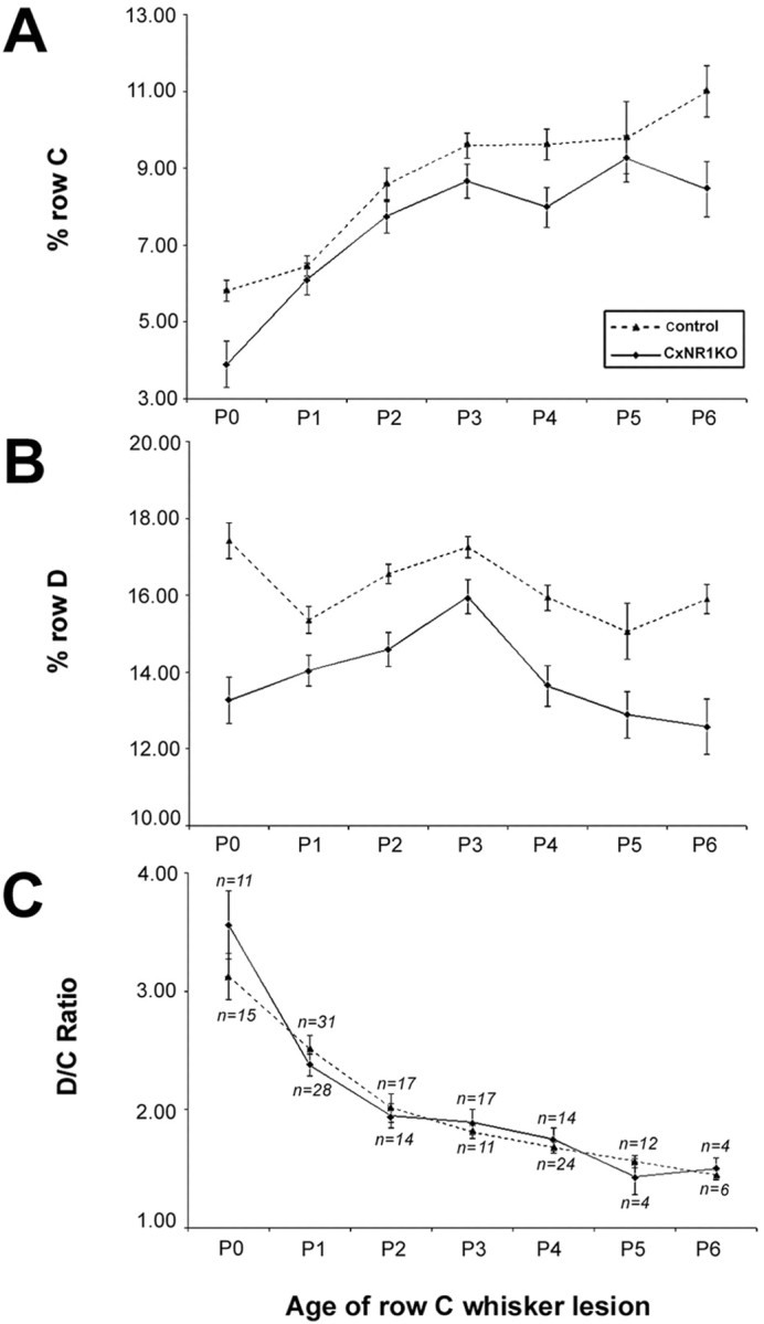
Quantification of lesion-induced critical-period plasticity in control and CxNR1KO barrel cortex. A, B, Analyses of normalized percentage row C and percentage row D areas at various ages of whisker lesions indicate that, despite the reduced areas of overall barrel rows in CxNR1KO S1 cortex (solid line), the shapes of the curves are similar, indicating that the TCAs respond similarly to the effects of peripheral lesions.C, The D/C ratios are indistinguishable for both genotypes and at different ages of row C lesion and follow a graded diminution until P3. Lesions placed after P3 are not effective in reorganizing TCAs, because the critical period of barrel plasticity has lapsed. n indicates number of animals. Error bars indicate SEM. Comparisons were made using ANOVA. For D/C ratios, there were no significant differences across all ages. Analysis of row D revealed significant differences between control (dashed line) and CxNR1KO cortices at all ages: P0,p < 0.000008; P1, p < 0.01; P2, p < 0.0003; P3, p < 0.04; P4, p < 0.0004; P5, p < 0.02; and P6, p < 0.002. Comparisons of row C between CxNR1KO and control cortices revealed significant differences for all ages except for P1, P2, and P3, probably because of significant contraction in row C area at these early ages. p values for other ages are as follows: P0, p < 0.0001; P4,p < 0.001; P5, p < 0.02; and P6, p < 0.001.