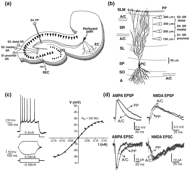
Fig 1.
Firing and subthreshold synaptic responses from CA3 pyramidal cells. (a) schematic diagram of hippocampal slice depicting the positions of the concentric bipolar stimulation electrodes (S1–S4). The recording electrode (REC) is in one CA3b pyramidal cell. Associational/commissural fibres (A/C) were activated from the str. radiatum (proximal (S1), medial (S2), distal (S3)). Perforant path fibres (PP) were activated from the str. lacunosum moleculare (S4) in area CA1. (b) reconstruction of a CA3 pyramidal neuron showing the typical soma location of CA3pc. CA3 pyramidal cells receive convergent excitatory synaptic inputs from other CA3 pyramidal cells and from the entorhinal cortex, via the A/C and the PP, respectively. The position of the electrodes for proximal, medial, and distal SR stimulations are shown. Abbreviations: A, alveus; SO, str. oriens; SP, str. pyramidale; SL, str. lucidum; SR, str. radiatum; SLM, str. lacunosum-moleculare. (c) Top left: adapting train of action potentials in response to a high intensity depolarizing current injection in a CA3pc. Bottom left: voltage changes in response to low intensity depolarizing and hyperpolarizing current injections in the same CA3pc. Right: I–V curve measured from the same CA3pc showing outward and inward rectification. (d) Representative AMPA and NMDA subthreshold EPSPs (top left and right) and EPSCs (bottom left and right) for A/C and PP inputs

