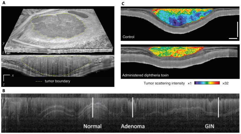
Figure 1.
Microstructural OCT imaging of tumours. (a) Using microstructural contrast, tumour tissue (an allotransplanted MCaIV mammary adenocarcinoma) can be differentiated from surrounding host subcutaneous and muscle tissues in a dorsal skinfold chamber model10. By defining the tumour margins, the three-dimensional tumour volume can be calculated. (b) Endoscopic microstructural OCT of azoxymethane-induced colorectal cancer in the mouse colon. High soft-tissue contrast and near histology resolution allows imaging of colonic epithelial microstructure12. The image shows the contrast at different tissue depths (shown in the vertical direction) versus distance along the colon (shown in the horizontal direction). Disease progression — from normal tissue to gastrointestinal intraepithelial neoplasia (GIN) to adenoma — can be monitored through hallmark modifications, such as the loss of tissue stratifications. (c) By quantifying scattering in OCT microstructural datasets, tumour viability can be monitored during cytotoxic interventions. Here, representative viability images of a LS174T human colorectal adenocarcinoma xenograft in a dorsal skinfold chamber model are presented two days following administration of diphtheria toxin (lower panel) or untreated (upper panel)10. Increases in scattering indicating significant loss of viability are evident in the diphtheria toxin administered animal. Transverse extent in a: 5 mm (x), 4.4 mm (y). Scale bars in c: 500 μm. Figure 1a and 1c are adapted from ref 10. Figure 1b is adapted from reference 12 with permission.

