Figure 1.

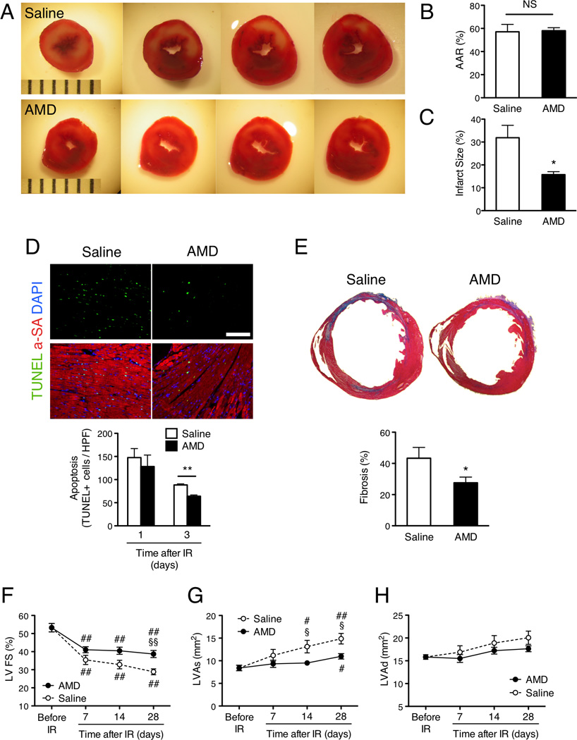
AMD3100 treatment improves cardiac function and reduces infarct size after IR injury. Mice were treated with saline alone or with 100 uL saline containing 125 ug AMD3100 after surgically induced IR injury. (A–C) AAR and infarct size were evaluated 3 days after IR injury via in-vivo microsphere perfusion and TTC-staining. (A) Viable tissue was stained deep red, and the infarcted region is colorless; scale=1 mm increments. (B) The AAR was identified by the absence of microspheres and presented as a percentage of the total left-ventricular (LV) area. (C) Infarct size was normalized to the size of the AAR and presented as a percentage. (D) Apoptosis was evaluated in TUNEL-stained sections of heart tissue from mice sacrificed 1 and 3 days after IR injury; scale bar=100 um. (E) Fibrosis was evaluated in Masson-trichrome-stained heart sections from mice sacrificed 28 days after IR injury, quantified as the ratio of the length of fibrosis (blue) to the LV circumference, and presented as a percentage. (F–H) Echocardiographic assessments of (F) LV fractional shortening (G) (FS), LV systolic area (LVAs), and (H) LV diastolic area (LVAd) were performed before IR injury and 7–28 days afterward; heart rates were maintained at 400–500 beats/minute via isoflurane inhalation. #P<0.05 and ##P<0.01 versus before injection; *P<0.05, **P<0.01, §Bonferroni-adjusted P<0.05 and §§Bonferroni-adjusted P<0.01 versus saline; NS, not significant. Panels B, C: n=3 per treatment group; Panel D: n=3–5 per treatment group; Panel E: n=6–9 per treatment group; Panels F-H: n=10 per treatment group at each time point.
