
Figure 2.
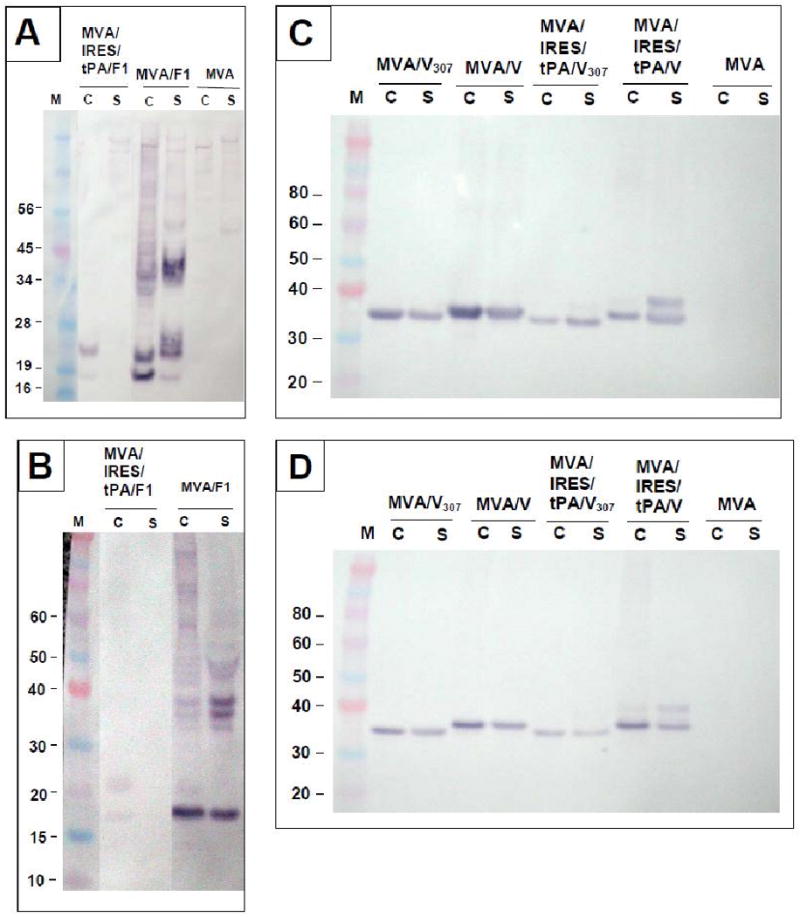
Western blot analysis of expression patterns from recombinant MVA-plague viruses.
Monolayers of CEF or Vero cells were transfected with recombinant MVA-plague viruses at MOI of 0.5pfu/cell. After 48 h post transfection cells were harvested and subjected to SDS-PAGE followed by western blot analysis as described in the methods. (A) F1 expression in CEF cell (c) and supernatant (s) fractions. (B) F1 expression in Vero cell (c) and supernatant (s) fractions. (C) V and V307 expression in CEF cell (c) and supernatant (s) fractions. (D) V and V307 expression in Vero cell (c) and supernatant (s) fractions.