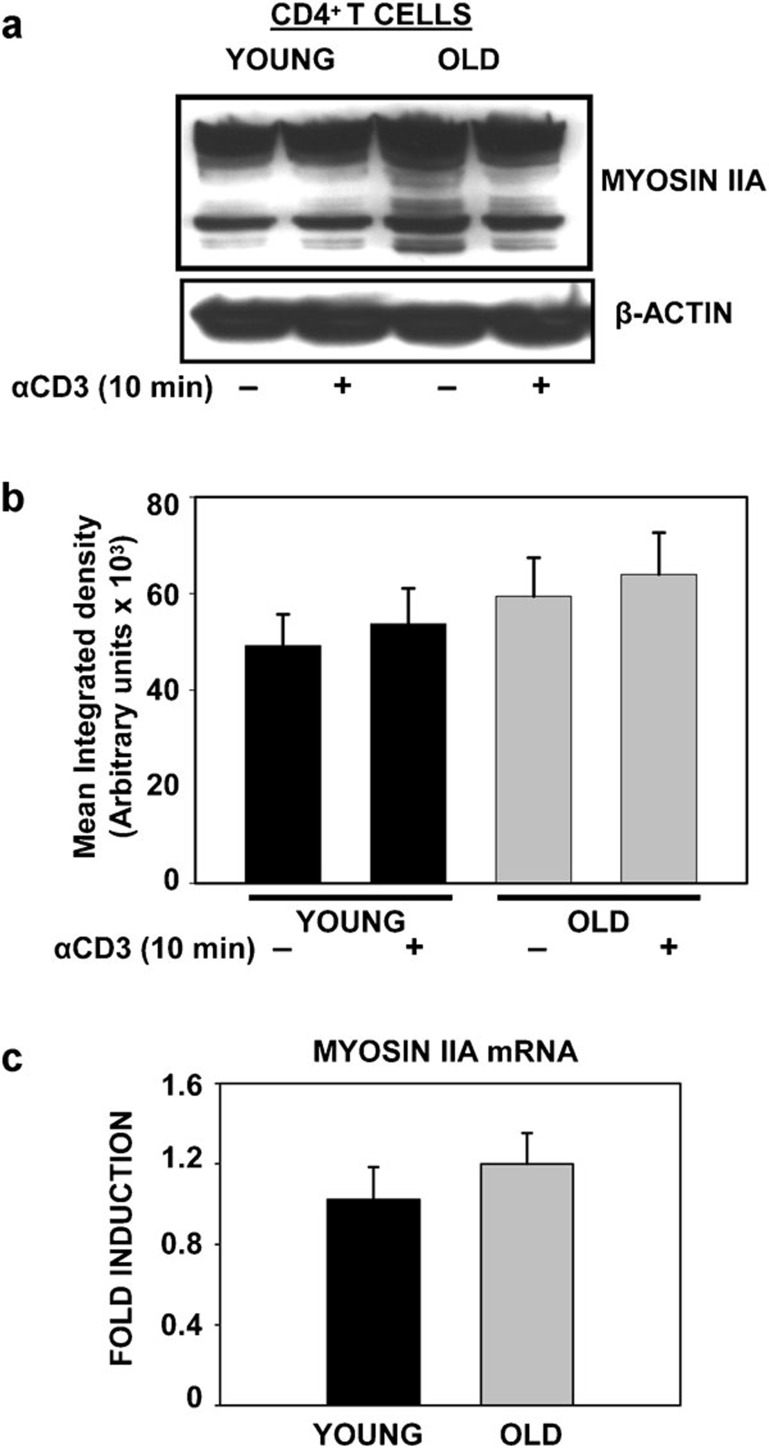
Figure 2.
Expression of non-muscle myosin IIA in CD4+ T cells from young and elderly donors. (a) Western blot of NMMIIA in CD4+ T cells from young and elderly donors either left untreated or activated with anti-CD3 beads for 10 min. β-actin was used as a control for equal protein loading. Representative data from one donor pair out of eight pairs tested are provided. (b) The specific band for myosin IIA protein was quantified by densitometry. The values represent the mean integrated density±s.e. obtained from a minimum of eight independent donor pairs. (c) qRT-PCR analysis of myosin IIA mRNA from total RNA collected from CD4+ T cells. The average data from 10 donor pairs are provided. β-actin and GAPDH were used as reference genes and for normalization. The data are presented as fold induction. GAPDH, glyceraldehyde-3-phosphate dehydrogenase; NMMIIA, non-muscle myosin IIA; qRT-PCR, quantitative PCR with reverse transcription.

