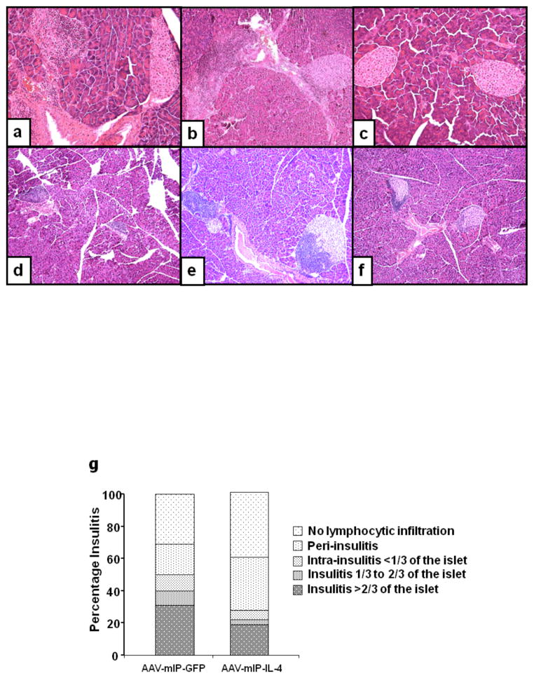
Figure 4. Histochemical analysis of infiltrating leukocytes in twenty week normo-glycemic mice.
Photomicrographs of H&E for infiltrating leukocytes in female 8 and 21 week normoglycemic female NOD mice showed mild to severe mononuclear cell infiltration in and around islets of saline and GFP treated mice (Figs. 4a–b; e–f), whereas the dsAAV8-mIP-IL-4 treated mice showed limited peri-islet mononuclear cell infiltration in significantly fewer numbers of islets (Figs. 4c, f). Figure 4g shows the insulitis score determined as mentioned in methods. Magnification 100X.

