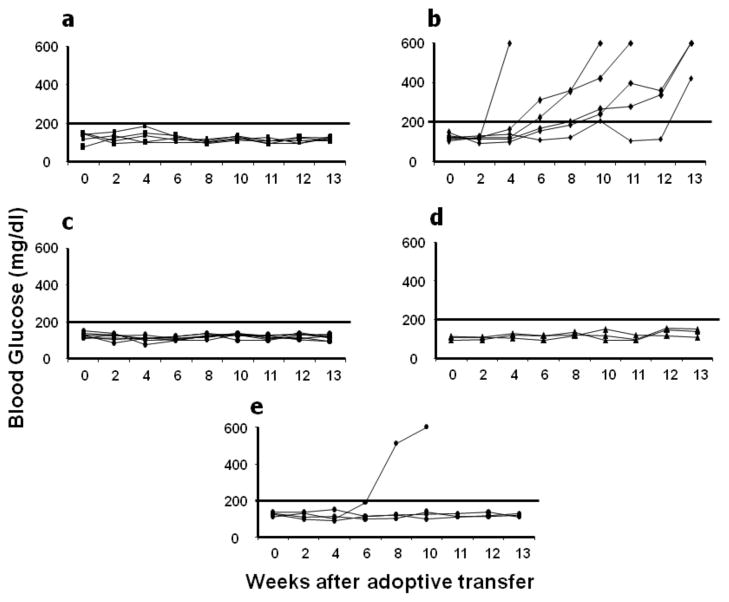
Figure 6. Adoptive transfer of diabetes in NODscid.
Splenocytes isolated from normal 21 week dsAAV-mIP-GFP or normal 21 and 40 week mIL-4 transduced mice were injected intra-peritoneally in to NODscid mice and blood glucose levels were monitored weekly. The NODscid mice which received splenocytes from GFP treated mice became diabetic (Fig. 6b), whereas the splenocytes from 22 and 40 week mIL-4 treated mice didn’t transfer the disease to NODscid (Figs. 6c–d). The NODscid mice receiving saline only also did not develop disease (Fig. 6a). To determine the suppressive effects of mIL-4 treated NOD mice splenocytes, co-adoptive transfer was performed as described in methods. Three of the 4 mice which received both splenocytes from mIL-4 and eGFP treated mice did not develop diabetes (Fig. 6e).

