
Figure 3.
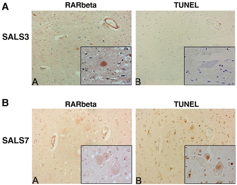
Immunohistochemical analysis of RARβ and TUNEL in human spinal cord. Serial lumbar spinal cord sections from SALS cases were immunostained for RARβ and TUNEL and representative cases are shown. Identical motor neurons were imaged as determined by anatomical hallmarks and location within the tissue. A: Motor neurons with intense nuclear RARβ immunostaining (A) lacked TUNEL staining (B). B: Motor neurons within the spinal cord without nuclear RARβ immunostaining (A) were TUNEL positive (B). Insets represent a high power magnification of each panel. Original magnifications: 200X; 400X (insets).